NORMAL ANATOMY OF BREAST

1.1  Introduction

A thorough understanding of the normal anatomical features of the breast is essential to develop skills in CBE and in different breast imaging techniques. Breast imaging using mammography, ultrasound, or magnetic resonance imaging (MRI) involves recognition of the morphological features of the normal and abnormal breast tissues. Knowledge of the gross anatomy and the microscopic architecture of the breast tissue enable the radiologist to recognize the underlying pathological changes when certain characteristics are observed during imaging studies.

1.1  Gross anatomy of the breast

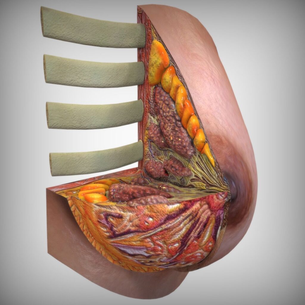
  • The adult female breast is situated between the second and the sixth ribs and extends horizontally from the edge of the sternum (the flat bone in the middle of the chest) to the mid-axillary line.
    • The breast overlies the deep fascia covering the pectoralis major muscle (pectoralis fascia), from which it is separated by loose areolar tissue containing small blood vessels and lymphatics. This loose connective tissue in the retromammary space gives rise to lucency in the mammogram.
    • The lucent area is known as the retromammary bursa or Chassaignac bag. The breast tissue often curves around the lateral free margin of the pectoralis major.
    • The axillary tail (tail of Spence) is a prolongation of the upper and outer quadrants of the breast towards the axilla. This tail passes under the axillary fascia (through the foramen of Langer) and may be mistaken for enlarged lymph nodes.

Each breast is divided into four quadrants, with the nipple and areola at the centre:

  • UOQ: Upper outer quadrant (superior and lateral)
    • LOQ: Lower outer quadrant (inferior and lateral)
    • LIQ: Lower inner quadrant (inferior and medial)
    • UIQ: Upper inner quadrant (superior and medial).

Skin and subcutaneous tissue

  • The skin of the breast contains the nipple and the areola.
  • The nipple is an erectile structure covered with thick pigmented skin, which also contains muscle. The orifices of the lactiferous ducts are located near the apex of the nipple.
  • The skin of the areola contains numerous modified sweat glands and sebaceous glands. These glands, also known as Montgomery tubercles, enlarge during pregnancy.
  • The areola contains involuntary muscles in the subcutaneous tissues that are arranged in concentric rings and radially.

The breast tissue underlying the skin is composed of:

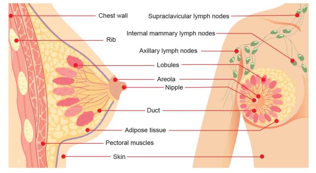
  • the glandular tissue (epithelial components), and
  • the adipose tissue supported by the fibrous connective tissue (stromal elements).

The glandular tissue (epithelial components)

The glandular component of an adult breast comprises 15–20 lactiferous lobes (segments) embedded in the fat and supported by the fibrous connective tissue. Each lobe is made up of 20–40 terminal duct lobular units (TDLUs), which are the functional units of the breast. Each TDLU comprises 10– 100 small sacs called glandular acini, which are connected to a terminal duct. The acini and the terminal ducts produce milk.

The terminal duct has an intralobular component and an extralobular component. Extralobular terminal ducts attach the lobule to the ductal system. Each lobule drains into a lactiferous duct, which converges to drain at the nipple–areolar complex. The lactiferous ducts dilate to form the ampulla (collection of lactiferous sinuses) as they converge to the nipple.

medicare
Breast Cancer – Global Overview – Updates

The International Agency for Research on Cancer (IARC) estimates that globally, 1 in 5 people develop cancer during their lifetime, and 1 in 8 men and 1 in 11 women die from the disease. These new estimates suggest that more than 50 million people are living within five years of a past cancer diagnosis. (IARC, Vol. 15) Ageing populations globally and socio-economic and lifestyle factors remain among the primary factors driving this increase. Breast cancer is the most commonly diagnosed cancer globally, which accounted for 1 in 8 cancers diagnosed and a total of 2.3 million new cases in both sexes combined in 2020 (GLOBOCAN 2020).The disease accounted for a quarter of all cancer cases in women in 2020, and the burden is growing in many parts of the world, particularly in transitioning countries. An estimated 685,000 women died from breast cancer in 2020, which corresponds to 16% or 1 in every 6 cancer deaths in women. By 2020, there were 7.8 million women alive who were diagnosed with breast cancer in the past 5 years, making it the world’s most prevalent cancer. There are more lost disability-adjusted life years (DALYs) by women to breast cancer globally than any other type of cancer. Breast cancer occurs in every country of the world in women at any age after puberty but with increasing rates in later life.

.

Figure 1: Age-standardized incidence rates in the world in 2020, all cancers, both sexes, all ages (Burden Global Age Standardized Incidence Rates in 2020)

A large geographic variation across countries and world regions exists, with incidence rates ranging from less than 40 per 100,000 females in some Asian and African countries, to over 80 per 100,000 in Australia/New Zealand, Northern America, and in large part of Europe. Smaller geographical variation was observed for mortality; however, transitioning countries continue to carry a disproportionate share of breast cancer deaths relative to transitioned countries. By 2040, the burden of breast cancer is predicted to increase to over 3 million new cases and 1 million deaths every year because of population growth and ageing alone.

The previously insufficient public health response to this development has led to the recent launch of the Global Breast Cancer Initiative (GBCI) in 2020 by the World Health Organization. By engaging global partners and coordinating sustainable efforts to improve outcomes, WHO and collaborators aim to reduce breast cancer mortality by fostering timely diagnosis and adequate treatment and patient management. The initiative focuses on steady escalation in the recognition of breast cancer as a public health priority by governments and stakeholders. The initiative is aimed to provide guidance to governments on how to strengthen systems for diagnosing and treating breast cancer, which in turn is expected to lead to improved capacities to manage other types of cancer. As a foundation to these efforts, a good understanding of global patterns and significant efforts to reduce the disease

burden is vital. Till now, significant disparities remain between higher and lower-income countries with the greatest relative increase in breast cancer incidence predicted to be observed in countries classified as “Low or Medium Human Development Index (HDI)” (includes Nepal). GLOBOCAN 2020-2040 prediction shows that low or medium HDI countries will have the greatest relative increases in cancer incidence and mortality by 2040.

Survival rates in high-income countries far exceed of the rates in low-income countries, with 5-year survival exceeding over 80% in most high-income countries as, compared to 66% in India and 40% in South Africa. The high out-of-pocket expenditure, lack of proper diagnostic and treatment facilities poses a significant threat to survival in low-income settings, like in Nepal.

medicare